Technologies Enhance Tumor Surgery
Helping Surgeons Spot and Remove Cancer

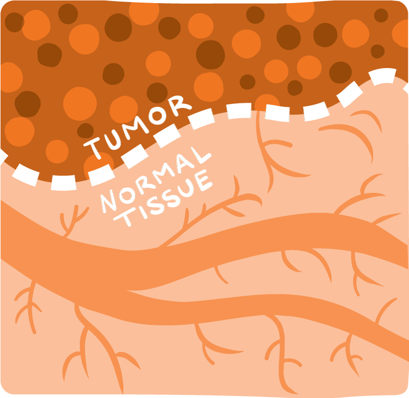
For surgeons, removing a tumor is a balancing act. Cut out too much and you risk removing healthy tissues that have important functions. Remove too little and you may leave behind cancer cells that could grow back into a tumor over time.
NIH-funded researchers are developing new technologies to help surgeons determine exactly where tumors end and healthy tissue begins. Their ultimate goal is to make surgery for cancer patients safer and more effective.
“Currently, surgeons view MRI and CT scans taken prior to an operation to establish where a tumor is located and to plan a surgical approach that will minimize damage to healthy tissues,” says Dr. Steven Krosnick, an NIH expert in image-guided surgery. “But once the operation has begun, surgeons generally rely only on their eyes and sense of touch to distinguish tumor from healthy tissue.”
Surgeons go through many years of training to understand the subtle cues that can distinguish tumor from normal surroundings. Sometimes the tumor is a slightly different color than healthy tissue, or it feels different. It might also bleed more readily or could contain calcium deposits. Even with these cues, however, surgeons don’t always get it right.
“In a lot of cases, we leave tumor behind that could be safely removed if only we were able to better visualize it,” says Dr. Daniel Orringer, a neurosurgeon at the University of Michigan.
In today’s operating rooms, Doctors who identify diseases by studying cells and tissues under a microscope. pathologists can often help surgeons determine if all of a tumor has been taken out. A pathologist may view the edges of the tissue under a microscope and look for cancer cells. If they’re found, the surgeon will remove more tissue from the patient and send these again to the pathologist for review. This process can occur repeatedly while the patient remains on the operating table and continue until no cancer cells are detected.
“Each time a pathologist analyzes tissue during an operation, it can take up to 30 minutes because the tissue has to be frozen, thinly sliced, and stained so it can be viewed under the microscope,” Krosnick says. “If multiple rounds of tissue are taken, it can greatly increase the length of the surgery.”
In the days following an operation, the pathologist conducts a more thorough review of the tissue. If cancer cells are found at the margins, the patient may undergo a second surgery to remove cancer that was left behind.
Orringer is part of a research team that’s testing a new technology that could help surgeons tell the difference between a tumor and healthy brain tissue during surgery. The team developed a special microscope with NIH support that shoots a pair of low-energy lasers at the tissue. That causes the chemical bonds in the tissue’s molecules to vibrate. The vibrations are then analyzed by a computer and used to create detailed images of the tissue.
From a molecular point of view, the components of a tumor differ from those in healthy tissue. This specialized microscope can reveal differences between the tissues that can’t be seen with the naked eye.
“Our technology enables us to get a microscopic view of human tissues without taking them out of the body,” Orringer says. “We can see cells, blood vessels, the connections between brain cells…all of the microscopic components that make up the brain.”
Orringer and colleagues developed a computer program that can quickly analyze the images and assess whether or not cancer cells are present. This type of analysis could help surgeons decide whether all of a tumor has been cut out. To date, Orringer has used the specialized microscope to help remove cancer tissue in nearly 100 patients with brain tumors.
Other researchers are taking different approaches. For example, Dr. Quyen Nguyen—a head and neck surgeon at the University of California, San Diego—has developed a fluorescent molecule that’s currently being tested in clinical trials. The patient receives an injection of the molecules before surgery. When exposed to certain types of light, these molecules cause cancer cells to glow, making them easier to spot and remove. The surgeon then uses a near-infrared camera to visualize the glowing tumor cells while operating.
Nguyen is also developing a fluorescent molecule to light up nerves. Accidental nerve injury during surgery can leave patients with loss of movement or feeling. In some cases, sexual function may be impaired.
“Nerves are really, really small, and they’re often buried in soft tissue or encased within bone. When we have to do cancer surgery, they can be encased in the cancer itself,” Nguyen says. The fluorescent molecule could help surgeons detect hard-to-spot nerves, so they can be protected. The nerve-tagging molecule is now being tested in animal studies.
Other NIH-funded researchers are focusing on ways to speed up cancer surgeries. Dr. Milind Rajadhyaksha, a researcher at Memorial Sloan Kettering Cancer Center, has developed a microscope technique to reduce the amount of time it takes to perform a common surgery for removing non-melanoma skin cancers.
Each year about 2 million people in the U.S. undergo Mohs surgery, in which a doctor successively removes suspicious areas until the surrounding skin tissue is free of cancer. The procedure can take several hours, because each time more tissue is removed, it has to be prepared and reviewed under a microscope to determine if cancer cells remain. This step can take up to 30 minutes.
The technique developed by Rajadhyaksha shortens the time for assessing removed tissue to less than 5 minutes, which greatly reduces the overall length of the procedure. Tissue is mounted in a specialized microscope that uses a focused laser line to do multiple scans of the tissue. The resulting image “strips” are then combined, like a mosaic, into a complete microscopic image of the tissue.
About 1,000 specialized skin surgeries have already been performed guided by this technique. Rajadhyaksha is currently developing an approach that would allow doctors to use the technology directly on a patient’s skin, before any tissue has been removed. This would allow doctors to identify the edges of tumors before the start of surgery and reduce the need for several pre-surgical “margin-mapping” biopsies.
There are many types of cancer surgeries, and researchers continue to work hard to develop better techniques. If you’re considering surgery to treat your cancer, you can find additional information at this NIH Cancer Surgery page.
NIH Office of Communications and Public Liaison
Health and Science Publications Branch
Building 31, Room 5B52
Bethesda, MD 20892-2094
Contact Us:
nihnewsinhealth@od.nih.gov
Phone: 301-451-8224
Share Our Materials: Reprint our articles and illustrations in your own publication. Our material is not copyrighted. Please acknowledge NIH News in Health as the source and send us a copy.
For more consumer health news and information, visit health.nih.gov.
For wellness toolkits, visit www.nih.gov/wellnesstoolkits.


