About Faces
The Biology of Face and Head Formation

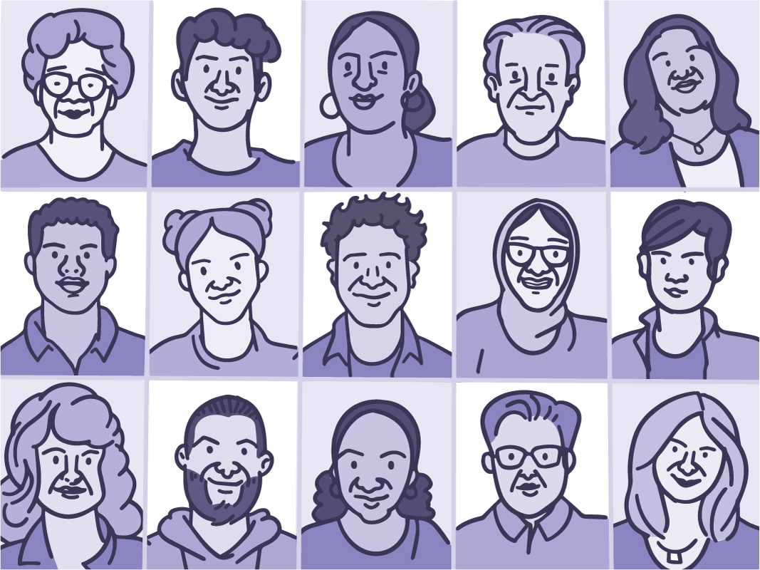
There’s a reason we can spot a friend in a crowd—humans are wired to focus on faces. We’re incredibly skilled at recognizing small differences in a face, like a square jaw, arched brows, or high cheekbones. The uniqueness of faces inspires artists and poets. It also enables facial recognition technology. The distinct features of each face help to define who we are.
“There’s a lot of information in a face,” says Dr. Seth Weinberg, who studies genes that affect the face and head at the University of Pittsburgh. “It’s how we connect with each other, understand emotions, and interpret social cues.”
Despite its importance, the underlying biology that creates each face remains unclear. And scientists are not yet certain what goes wrong to cause birth defects of the head and face. These are called craniofacial disorders. They can make it hard to eat, hear, speak, see, and breathe. Cranofacial disorders can also harm the growing brain.
NIH-funded researchers are working to unravel the mysteries behind how the head and face develop. Their findings could not only help prevent or treat craniofacial disorders, like cleft lip and palate. They could shed light on the function and development of other body parts, since the head and face include many nerve cells, bones, immune cells, and more.
Molding the Face and Head
One way to decipher the underlying biology of the face and head is to gather data—lots of it. Scientists analyze genetic information, take images of people’s faces, and collect other biological information from both humans and animals. And they share this data with other scientists to enable discoveries.
So far, researchers have linked over 300 areas of our DNA to facial features like nose height, eye width, and chin shape. In one study, Weinberg and colleagues analyzed images of the head from more than 6,000 children. This helped them uncover previously unknown sets of genes that can affect the shape of the human head. These findings, in turn, could shed light on genetic disorders that affect the skull.
But genes alone don’t tell the whole story. Even identical twins with the same genes don’t always look exactly alike.
“From our research, our genes only explain about 14% of the variation in facial features,” Weinberg says. Our age, diet, environment, exposure to chemicals, and many other factors can mold the shapes of our faces before and after birth.
Tailoring Treatment
While scientists haven’t yet pinpointed all the factors that affect our faces, they do know that when craniofacial disorders arise, they generally begin before birth. These disorders occur when bones, nerves, and tissues in the face and head don’t form properly as a baby is growing in the womb.
For example, cleft lip and palate is a birth defect that arises around the second or third month of pregnancy. It occurs when the right and left sides of the lip, the roof of the mouth (called the palate), or both don’t join all the way. This creates a gap, or cleft.
“Cleft lip and palate is the most common craniofacial disorder. Instead of a continuous lip, there is a notch or there’s a defect that extends up into the nose, so the lip is in two segments,” explains NIH’s Dr. Janice Lee, who specializes in surgery for the face, head, neck, and jaw. “Typically, we can identify it while the baby is growing in the womb or at birth.” With 3D imaging techniques, doctors can now assess cleft disorders before birth and begin to plan repairs.
Newborns with cleft lip or palate are often referred to a team of surgeons, dentists, geneticists, pediatricians, and speech therapists for care. These experts may follow their patients from birth to adulthood, repairing the cleft and guiding recovery. Cleft lip and palate can affect a child’s oral health and social well-being. The goal is to tailor care for each patient and lessen the disorder’s impact on their lives. With treatment, most children with cleft lip or palate do well and lead a healthy life.
Finding New Options
Surgeries for the face and head can be complex and tough on the body. Even after surgery, some children may have trouble eating, breathing, and speaking. Scientists are continuing to develop new surgical techniques to help patients speak better and improve how their faces look. Others are creating computer programs and artificial intelligence tools to plan surgery for cleft lip or palate.
Researchers are also exploring ways to fix craniofacial disorders while reducing surgical procedures. Dr. Yang Chai at the University of Southern California aims to find ways to correct a craniofacial disorder called craniosynostosis.
Normally, a newborn’s skull bones are separated by flexible joints that make space for the brain to grow. But in babies with craniosynostosis, the joints close too soon. This can change a baby’s head shape and brain growth.
“During my surgical training, I performed surgeries to fix these conditions but couldn’t explain to parents why their kids had them,” Chai says. “This became a strong drive for me to better understand the disease and find a better solution for these patients.”
Chai and his colleagues are testing ways to grow more tissue between the skull joints in young mice with craniosynostosis. The researchers are using stem cells to fix the skull shape and reverse learning and memory problems in mice. Stem cells are special cells that can turn into many other types of cells, including bone, skin, and muscle. Their findings suggest that stem cell therapy may one day be a less invasive treatment for craniofacial disorders.
Predicting who might be more likely to have craniofacial disorders is another area that scientists are excited about. “If we can understand who’s at greatest risk or which families are at risk, we can do things that could potentially prevent these conditions from occurring,” Lee says. “We’re not there yet. But prediction and early treatment is really something we’re all working toward.”
NIH Office of Communications and Public Liaison
Health and Science Publications Branch
Building 31, Room 5B52
Bethesda, MD 20892-2094
Contact Us:
nihnewsinhealth@od.nih.gov
Phone: 301-451-8224
Share Our Materials: Reprint our articles and illustrations in your own publication. Our material is not copyrighted. Please acknowledge NIH News in Health as the source and send us a copy.
For more consumer health news and information, visit health.nih.gov.
For wellness toolkits, visit www.nih.gov/wellnesstoolkits.


